Internal Anatomy
Pyrazus ebeninus are unlike other gastropods in that they are not filter feeders, as exemplified by the lack of food groove on pallial floor and absence of endostyle at the base of the ctendium (Strong et al. 2011). The hypobrancial gland is well developed with pendulous folds for producing mucous indicating that rely strongly on their muscular foot for movement (Fig. 1, Pontarotti 2010). The radular sac inside of the buccal cavity is short and curves slightly under the oesophagus containing the radula used for feeding (Fig. 2; Maggenti & Gardne, 2005). Their central tooth basal plate present in the radula is wide with straight lateral sides of the central tooth. There are two denticles present with basal plate that extend beyond cutting edge, similar to other Potamididae. Unlike other gastropods, their central tooth glabella and basal extension is absent (Strong et al. 2011). However, the lateral tooth lateral extension is approximately twice the length of the cutting edge. Due to that P. ebeninusare detrital, non carnivorous feeders, the foregut has small, paired, non-modified chitinous jaws that partially bilayered at anterior end of dorsal folds. These organisms also have a complex salivary gland that is massive without common lumen passing through the circum-oesophageal nerve ring (Fig. 2, Strong et al. 2011). Like most other gastropods, P ebeninus has the presence of a hemocoel due to its compact body structure. This hemal system consists of the generalized structure of a heart, aorta, blood flow with blood vessels (Fig. 2). The osphradium chemoreceptive sense organ is narrow and located on the surface of the organism to monitor water currents (Wedemeyer & Schild, 1995). It is considered to be slightly shorter than the broad shaped ctendium and a short efferent ctenidial vein is found between the gills and the pericardium (Strong et al. 2011).
A typical Gastropod gut contains a mouth, buccal cavity, esophagus, stomach, intestine, anus, and rectum (Ruppert et al. 2004). There are no buccal pouches in the buccal cavity, although there is an esophageal gland located in the submucous material above the buccal mass (Fig. 2). The mid gut contains an S-shaped marginal fold and the oesophageal aperture is at or near tip of the recurved segment of this fold. The sorting area of the organism contains a system of ciliated grooves that is rectangular shaped with tapering occurring to create a curved posterior tip, while the left margin is seen as a large bulge (Strong et al. 2011). The marginal fold terminates at tip of sorting area with the accessory marginal fold segment located along the left side of sorting area. This configuration causes the formation of a deep pocket overhanging posterior tip. The glandular pad is a large, long and narrow mound with a digestive gland duct located open to left side of glandular pad (Fig. 1). The cresentic ridge is double and curved closely adhering to the glandular pad. The ridge proximal edge begins at digestive gland duct and the distal end is attached to the right side of the glandular pad behind the gastric shield (Strong et. al 2011).
The mid-gut is u-shaped folded and is limited to the visceral mass (Fig. 3). All digestion processes occurring are to some degree extracellular in the stomach with the enzymes being produced from the either the salivary glands, esophageal pouches, or digestive ceca (Ruppert et al. 2004). Secondarily, the digestive ceca is also used for absorption and intracellular digestion in some gastropods. A large portion of freshwater taxa made modifications to the kidney and have created a separation of the dorsal concentration of excretory tissue (Stronget al. 2011). This modification creates a more ventral bladder with the septum perforated by a small aperture. This formation allows communication between the kidney and the bladder (Strong et al. 2011). The bladder holds variable amounts of excretory tissue and communicates with the mantle cavity through the nephropore. The nephropore, an opening on the antennal gland, may be simple or subdivided by an incomplete septum and is not documented which form occurs in this organism (Strong et al. 2011). Excretion in marine gastropods occurs through diffusion across the gills and body surface ultimately excreting ammonia and losing nitrogen (Ruppert et al. 2004). Due to that many intertidal species can vary between ammonotelic or ureotelic depending on when the tide is in or out, it is difficult to determine which excretion characteristic P. ebeninus has. The paracardial glands present are used in the foramtion of urine consistent with other molluscs. The paracardial gland produces an ultrafiltrate in the paracardial cavity that flows through the renoparcardial duct into the nephridium, where the epithelium converts the primary urine into final urine to be released into the mantle cavity through the nephridiopore (Ruppert et al. 2004). Despite complex kidneys seen in other taxa, P. ebeninus have no kidney bladder, but rather a simple kidney opening contrary to the diagram in Figure 4. The nervous system of gastropods consists of a bundle of several ganglia forming a brain-like structure, the central ganglia. The P ebeninus nervous system exemplifies cebral ganglia that has a short commissure, indicating that the organism has direct contract or slight constriction that is less than half as long as an individual cerebral ganglion (Strong et al. 2011). These organisms, like other gastropods, have a epiathroid nervous system meaning that the pleural ganglia lies close to the cerebral ganglia (Chase 2002). Zygosis neural connections occurs between the right pleural and sub esophagealganglia located close to left pleural ganglion. The supra oesophageal ganglion is widely separate from right pleural and numerous calciferous statoconia located in the gelatinous membrane of the acoustic maculae (Strong et al. 2011).
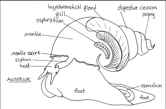
Figure 1: Generalized diagram of the internal anatomy of a marine gastropod. All of the above descriptive features are present in P. ebeninus organisms.
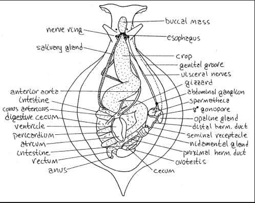
Figure 2: A detailed diagram of the internal anatomy of a marine gastropod. Some of the organs listed are not present in P. ebeninus as described above. In addition, it should be noted that the reproductive system seen in this figure exhibits a hermaphroditic lifestyle with hermaphroditic ducts, whereas P. ebeninus experiences gonochoristic reproduction with only one sexual reproduction organ.
Figures 1 & 2 were described by Fox 2001 redrawn from Kandel 1979
Source: http://lanwebs.lander.edu/faculty/rsfox/invertebrates/aplysia.html
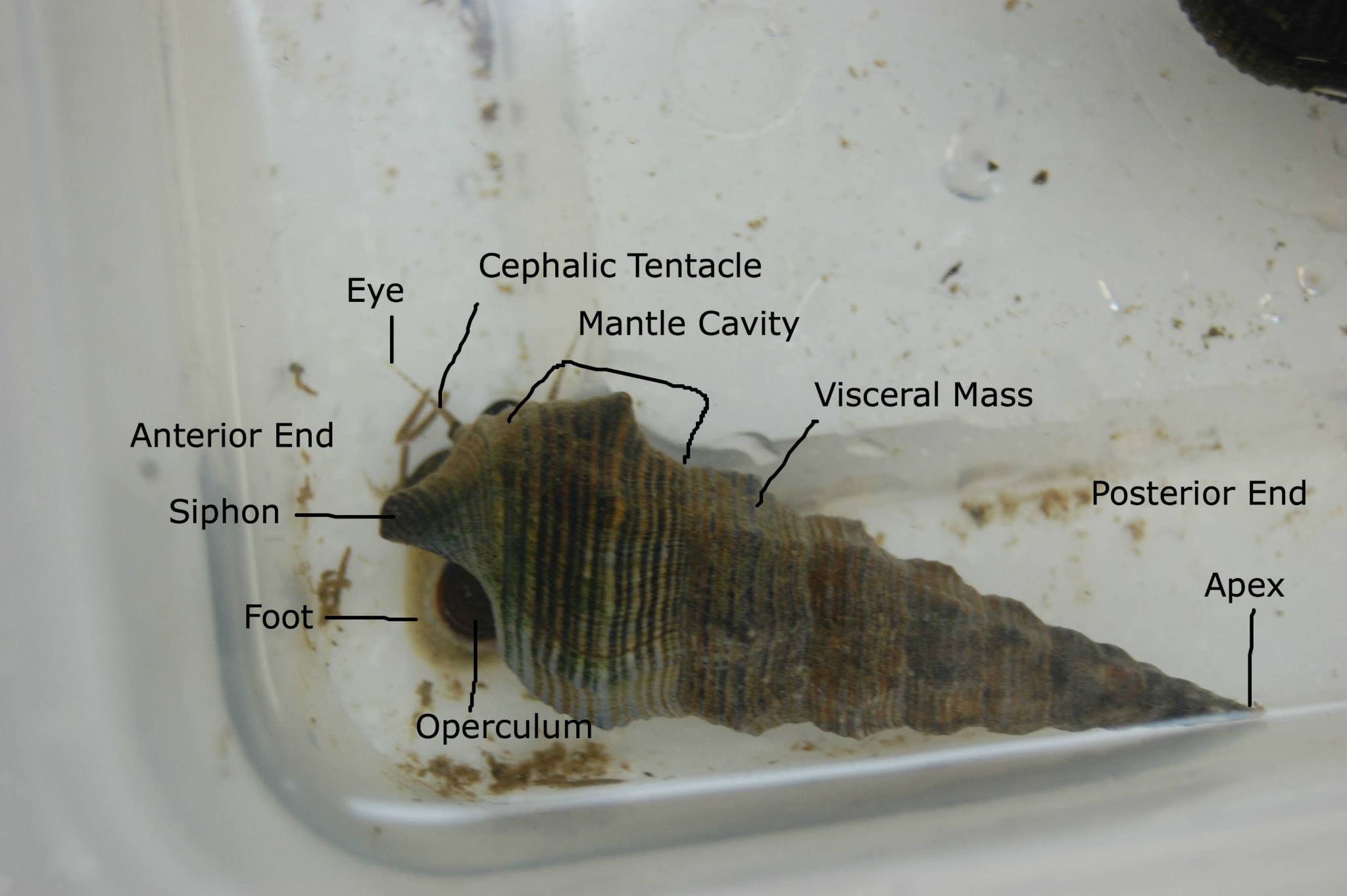
Figure 3: Generally labeled diagram of the external and internal anatomy of a P. ebeninus specimen in sample tray.
|